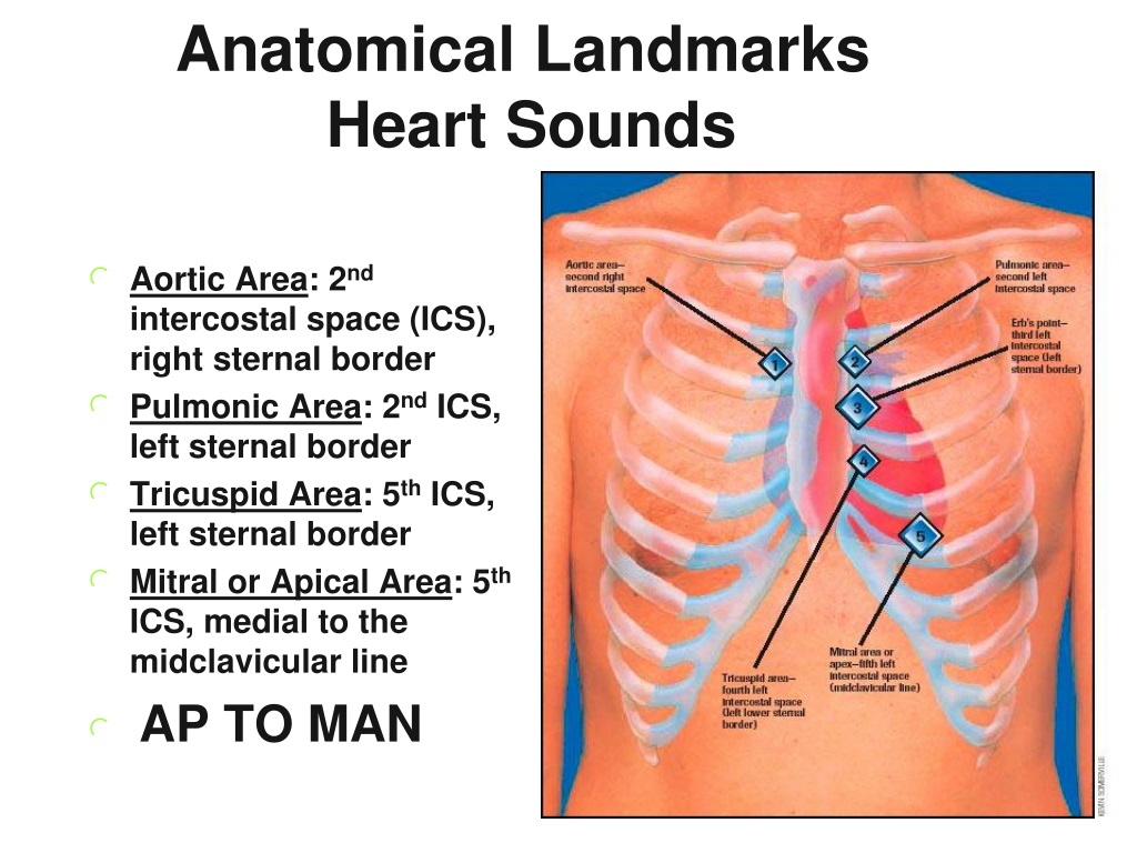
Diastole: The period between S2 and the beginning of the next heart beat (S1). Systole: The period between S1 and S2, when the ventricles contract. S4 is an atrial gallop, produced by the atria forcefully pushing blood into a stiff ventricle. S3 is a ventricular gallop, a low-pitched sound that can follow S2. S2: The secound heart sound, caused by closing of the aortic and pulmonary valve. S1: The first heart sound, a low-pitched sound caused by the closing of the mitral and tricuspid valve. Heart sounds can include multiple sound components. Links to our lessons, guides and quizzes are found in the 'Quick Links' box to the left. This includes gaining an understanding of cardiac rate and rhythm, conditions of the valves and possible anatomical abnormalities such as congenital defects. On this website, we provide lessons, reference guides and quizzes for cardiac auscultation of murmurs and other heart sounds. This "lub-DUB" sound changes, often with additional sounds being heard. Stethoscopes are used to listen to heart murmurs.Ī normal heartbeat sounds like "lub-DUB", which are the sounds of your heart valves closing. However, other abnormal heart murmurs indicate Innocent murmurs frequently resolve without treatment. They can be found in infants or develop later in life. To learn more about these, click here.Heart murmurs are sounds produced by turbulent blood flow, particularly from the heart's valves. ventricular systolic failure)Īssociated with stiff, low compliant ventricle (e.g., ventricular hypertrophy ischemic ventricle)īesides these four basic heart sounds, other sounds such as murmurs can be heard. Normal in children in adults, associated with ventricular dilation (e.g. This sound is usually associated with a stiffened ventricle (low ventricular compliance), and therefore is present in patients with ventricular hypertrophy, myocardial ischemia, or in older adults. The fourth heart sound ( S 4), when audible, is caused by the vibration of the ventricular wall during atrial contraction. This sound is normal in children, but when heard in adults it is often associated with ventricular dilation as occurs in systolic ventricular failure. The third heart sound ( S 3), when audible, occurs early in ventricular filling, and may represent tensing of the chordae tendineae and the atrioventricular ring, which is the connective tissue supporting the AV valve leaflets. 
The timing of S 2 splitting changes depending on the phase of respiration, body posture, and certain pathological conditions.

S 2 is produced by the closure of the aortic and pulmonic valves at the beginning of isovolumetric ventricular relaxation, and the sound is physiologically split because aortic valve closure normally precedes pulmonic valve closure. S 1is normally slightly split (~0.04 sec) because mitral valve closure precedes tricuspid valve closure however, this very short time interval cannot normally be heard with a stethoscope, so only a single sound is perceived.

S 1 is caused by closure of the mitral and tricuspid valves at the beginning of isovolumetric ventricular contraction.

The most fundamental heart sounds are the first and second sounds, usually abbreviated as S 1 and S 2.


 0 kommentar(er)
